 Writers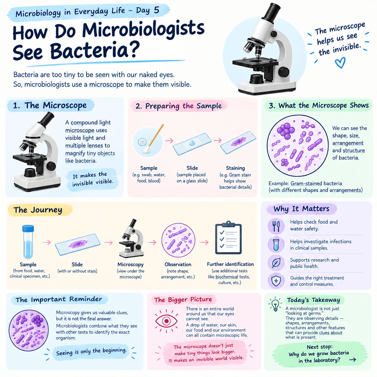
Microbiology in Everyday Life — Day 5
How Do Microbiologists See Bacteria?
Yesterday, we talked about bacterial shapes cocci, bacilli, spirals and vibrios.
But there is one obvious question:
How do microbiologists know what bacteria look like when bacteria are too small for our eyes to see?
The answer is one of the most important tools in the laboratory:
The microscope.
Your eyes have limits
Look at your hand.
You can see your skin, your fingers and even tiny marks on your palm. But if you wanted to see an individual bacterium sitting on that skin, your eyes would not be enough.
Bacteria are microscopic organisms. That means we need an instrument capable of making very small objects appear large enough to observe.
That is where microscopy comes in.
A compound light microscope, for example, uses visible light and multiple lenses to produce an enlarged image of a specimen. It is one of the common microscopes used in laboratory work.
So what does a microbiologist actually do?
It is not simply a matter of putting a sample under the microscope and looking.
A specimen may first need to be prepared on a glass slide. Depending on what the microbiologist wants to investigate, the specimen can undergo different preparation or staining procedures.
One important example is the Gram stain.
Gram staining helps microbiologists observe differences in bacterial cells and is routinely used as part of bacterial identification. Laboratory professionals can examine the stained cells under a microscope and note features such as their shape and arrangement.
So remember the journey:
Sample → Slide → Microscopy → Observation → Further identification
Why can't the microscope tell us everything?
This is where people sometimes misunderstand microbiology.
Suppose you look through a microscope and see several purple, round cells arranged in clusters.
That observation gives you useful information.
But you cannot simply say:
“I have identified the exact bacterium.”
Not necessarily.
Microscopy gives important clues, but microbiologists often combine microscopic appearance with other information, such as colony characteristics, biochemical tests and other laboratory methods, before identifying an organism.
In other words, seeing is only the beginning.
The microscope is more than a school laboratory instrument
Think about a microbiology laboratory.
Samples may come from clinical specimens, food, water or other environments. Being able to visualize microorganisms can help laboratory professionals investigate what is present.
The microscope is therefore not just something students use during practical classes.
It is an important laboratory tool for observing organisms that would otherwise remain invisible to us. CDC laboratory training specifically identifies microscopy as part of the skills used to identify microorganisms.
And there is something beautiful about this
There is an entire world around us that our eyes cannot see.
A drop of water can contain microscopic life.
The surface of our bodies can support microbial communities.
A laboratory sample can contain organisms that are completely invisible until a microscope reveals them.
Microbiology gives us a way to investigate that hidden world.
The microscope doesn't just make tiny things look bigger.
It makes an invisible world visible.
Today's takeaway
The next time you see a microbiologist looking through a microscope, remember that they are not simply “looking at germs.”
They are observing details that the naked eye cannot see shapes, arrangements, structures and other features that can provide clues about what is present.
And even then, the investigation may not be over.
Because in microbiology, seeing an organism is one thing; knowing exactly what it is requires evidence.
Next stop: What happens when bacteria are placed on a culture plate? Why do microbiologists grow bacteria in the laboratory?